Berta Domènech, Alvin T. L. Tan, Hans Jelitto, Eduardo Zegarra Berodt, Malte Blankenburg, Oliver Focke, Jaclyn Cann, C. Cem Tasan, Lucio Colombi Ciacchi, Martin Müller, Kaline P. Furlan, A. John Hart, Gerold A. Schneider
Advanced Engineering Materials (2020) 22, 2000352
https://doi.org/10.1002/ade…
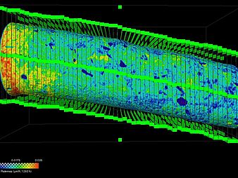
![[Translate to English:]](/fileadmin/_processed_/d/0/csm_XRM_Nils_c7a1a6f19a.png)
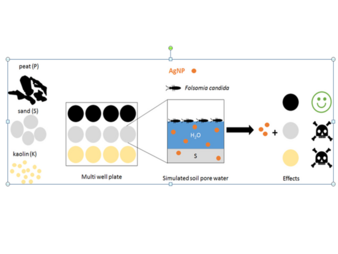
![[Translate to English:]](/fileadmin/_processed_/7/8/csm_Dilissen_etal_2021_8f8a0326b9.jpg "[Translate to English:]")