Extracellular Recordings

Neurons communicate via small electric pulses, called action potentials or spikes. In general, a neuron generates a spike (or many of them) when it receives sufficiently strong input at its synapses. Neuronal activity results in changes in the composition of ions in extracellular space and thereby in local variations of the electric field, which can be measured. Such extracellular recordings provide access to the activity of single cells and allow the investigation of communication between individual neurons and groups of neurons with very high spatio-temporal resolution. This is key to understand the neuronal mechanisms subserving cognitive functions, as e.g. attention-dependent improvement of neuronal response selectivity and signal-to noise ratio, coordination of activity in neuronal ensembles, and communication within and across brain areas (e.g. Grothe et al., 2012; Drebitz et al., 2018; Galashan et al., 2013; Schledde et al., 2017).
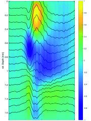
Onset response of visual neurons to a stimulus which is presented on a screen.
Die Abbildung zeigt die neuronale Aktivität innerhalb einer kortikalen Kolumne. Um diese aufzunehmen wurde eine Multi-Kontakt Elektrode verwendet, welche im Abstand von 100 µm das elektrische Feld misst. Das Bild zeigt die Aktivität der Neurone innerhalb der Kolumne während ein Stimulus im rezeptivem Feld der Neurone gezeigt wird (Onset). Neurone in tieferen Schichten regieren etwas früher auf die eintreffenden Signale (hier Blau). Diese Neurone befinden sich in der "Input" - Schicht und liefern ihrerseits Signale an Neurone in höheren (hier gelb) aber auch tieferen (grün) Schichten.
The map shows the visual area of a neuron to which it responses. The center of gaze is slighlty above the responsive region
Das Bild zeigt das Antwortverhalten eines Neurons im visuellen Areal 1 (V1). Die Aktivität des Neurons steigt stark an, wenn sich ein Objekt innerhalb des rezeptiven Feldes (hier rot gefärbt) befindet. Befindet sich das selbe Objekt an einem anderen Ort innerhalb des Sehfeldes, reagiert das Neuron wenig bis gar nicht (blau gefärbt).
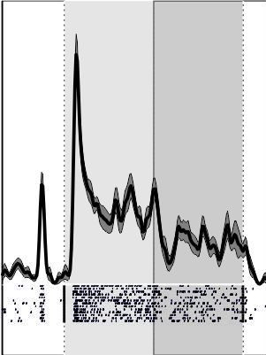
The spiking activity of neurons located in the visual middle temporal area. These neurons are very sensitive to motions and changes in speed. The shown trace depicts their activity (firing rate) in response to a moving grating, which accelerated in speed.
Aufgenommene neuronale Aktivität (Feuerrate, Aktionspotentiale pro Sekunde) eines Neurons im visuellen Areal MT (middle temporal). Neurone in MT sind besonders reaktiv für Bewegungen. Die gezeigte Aktivität ist die Antwort des MT Neurons auf ein sich bewegendes Gitter (moving grating), welches sich im rezeptiven Feld des Neurons befindet. Die Bewegung des Gitters beschleunigt sich dann, worauf das Neuron mit einer starken Erhöhung der Feuerrate reagiert (Transiente). Die Feuerrate sinkt daraufhin wieder ab, bleibt jedoch auf einem höheren Niveau als während der langsameren Geschwindigkeit zuvor.