Research

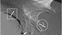
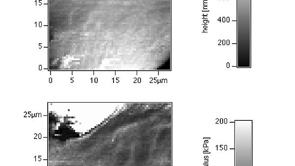
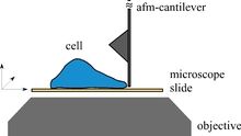
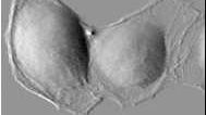
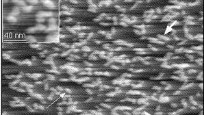
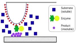
The research in the Radmacher lab focuses on the investigation of biological and biophysical processes. Examples are cell dynamics, cell mechanics, cell migration, cell division, activity of single enzyme molecules and enzymatic nanolithography. The most important tool for these investigations is the atomic force microscope (AFM). The AFM allows to investigate biological samples without any chemical fixation under physiological conditions combined with a high accuracy in positioning a very small tip and a high sensitivity in measuring and applying forces. This combination allows for instance to investigate biological processes from a cellular level down to single molecules. Although the AFM has been invented as an imaging device it has found recently several applications, where it is used to either to measure or to apply locally small forces on a sample. To learn more on AFM please click here.

Cellular Dynamics

Activation

One field of interest is the mechanics of cellular dynamics. In particular we are interested in the role of the cytoskeleton in cell migration and cell division. 

Cell Migration

Cell migration is a fundamental process in many biological processes, e.g. wound healing and metastasis. Although the mechanism generating the protrusion force at the leading edge of a cell is still unclear, the emerging image is that polymerization of actin plays an important role. In fact, it may actually generate the force itself by the polymerization itself. The physics behind it is covered in modles like the polymerization ratchet, developed by Mogilner and Oster, and its more elaborate variants like the elastic polymerizing ratchet and others.

We have investigated the mechanical properties of migrating cells and recently have designed an instrument to directly measure the forces of migrating keratocytes.

Cellular Mechanics

The AFM can be used to determine the mechanical properties of soft samples, e.g. cells. To learn more you may want to look into this short introduction.

The mechanical properties of cells are determined mainly by the actin cytoskeleton, which is (grossly simplified) a cross-linked polymeric network. Therefore they are very important in many cellular processes like cell migration and cell division.
The mechanical properties of the cytoskeleton can be probed by locally pressing onto the cell with a defined force and detecting the resulting indentation of the cell. Technically this is done by taking a force curve and analyzing it off-line. Thus maps of the elastic properties of the cell can be obtained.

An animated version of topographic images at increasing loading force reconstructed from another force map can be found here

Measuring protrusion forces

For measuring the forces at the leading edge of a protruding lamellipodium we have placed an AFM cantilever perpendicular to the sample in front of a cell. By video microscopy the deflection of the cantilever can be analyzed.
For more information, you may want to look at our publications.

Cell Division

It is unclear whether cell division is driven by cortical relaxation outside the equatorial region or cortical contractility within the developing furrow alone. To approach this question, a technique is required that can monitor spatially-resolved changes in cortical stiffness with good time resolution. We employed atomic force microscopy (AFM), in force-mapping mode, to track dynamic changes in the stiffness of the cortex of adherent cultured cells along a single scan-line during M phase, from metaphase to cytokinesis. Video microscopy, which we used to correlate the AFM data with mitotic events identified by light microscopy, indicated that the AFM force-mapping technique does not perturb dividing cells. Here we show that cortical stiffening occurs over the equatorial region about 160 seconds before any furrow appears, and that this stiffening markedly increases as the furrow starts. By contrast, polar relaxation of cells does not seem to be an obligatory event for cell division to occur.

Activity of Single Enzyme Molecules

The combination of nanometer precision in positioning and a force sensitivity of around 10 pN made it possible to investigate processes on the level of single molecules.
We have shown that the activity of single enzyme molecules can be probed by AFM. This opens a new field in studying the kinetics and characteristics of biological events of single molecules. The first studies used lysozyme as the system of investigations. However, there are many more molecules which seem to be ideal candidates for this type of experiment: urease, hexokinase, glucose-oxidase and others. It will be very interesting to "feel" differences of the activity of these molecules and thus develop a mechanical fingerprint for each enzyme. Undoubtedly new insights in the functioning of the molecular machinery will emerge from these studies. Along this line, we will try not only to observe the active motion of these molecules during their function, but it could also become possible to monitor spontaneous fluctuation in shape of the molecules, in other words to observe protein motion.We could also demonstrate that it is possible to investigate a single protein molecule for very long times (more than half an hour, see: Biophysical Journal (1996) 70, (5) 2421-2431). To hear sound tracks of different enzymes please click here.

Enzymatic Nanolithography

By immobilizing a few enzyme molecules ontop the tip of an AFM we were able to locally modify a suitable support. This process, called enzymatic nanolithography, has the potential to become a general tool for structuring surfaces on a nanometer scale.  For more information see our publication in Nanoletters.