
...a non-destructive method to explore the spatial distribution of matter
ProCon CT-ALPHA

Hydrothermally altered sandstone
Width ca. 9 mm

Ammonit
Width ca. 60 mm

Fishbones
Width of imaged structure ca. 45 mm

Submarine massive sulfur deposit
Width of micro core ca. 25 mm

Submarine volcanoclastic material
Width of sample ca. 1.5 mm

Human tooth
Height ca. 20 mm

Foraminifera with pyrite filling
Height of largest rendering XX mm

Lightweight aggregate
Width of 2D slice ca. 13 mm
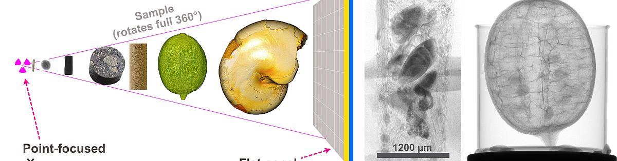
The Scanner
Our CT-ALPHA is equipped with a X-RAY WorX source (XWT-190-TCHE, 190 kV) that offers three different foci modes. Both a high-energy and a high-resolution target (W on diamond window) are in use. We can analyse both mm-sized samples with high resolution, and cm-sized samples with high energy. The Vieworks detektor (flat panel sensor VXTF-2532EAH, 2560 x 2048 Pixel, pixel pitch 124 μm) can be operated in double view mode.
The method
The method is based on the detection and localisation of the degree of attenuation of the incident X-rays in the sample. The attenuation of X-rays by matter depends on both the chemical elements it consists of as well as the material density. The information about the varied X-ray absorption is encoded as grey values in a stack of black-and-white images. Each image consists of volumetric pixels and contains true volumetric information.
Sample types and preparation
X-ray micro tomography can characterize all materials that are transparent for X-rays, e.g. silicates, carbonates, sulfates etc. The sample surface does not have to be prepared in advance. In our lab we analyze rocks with silicate, carbonate and sulfide minerals, volcanic pumice, foraminiferae, bivalves, organic samples like fish or rat bone, and more.
moreSample size and -geometry
In general, samples of any shape can be investigated, however a cylindrical shape is most favorable. Sample size may range from a few millimeters to several centimeters. Most important for a successful scan is a sufficient transmission of the sample for X-rays. It is possible to scan a selected sector of a sample (e.g, a micro core in a X-ray-transparent core holder).
What resolution can I expect?
Relevant for the attainable resolution is the position of the sample in the cone beam geometry between the X-ray point-focus source and the detector, which determines the magnification. In general, small samples (1-2 mm) can be characterised with high resolution (<1 - 3 μm per voxel), and larger samples with lower (20-40 μm per voxel).
moreWhat kind of result do I get?
The result is the reconstructed density distribution within the object in the form of a digital image stack or as image volume, which can be downloaded from the lab server. A storage medium will not be provided. In addition, the transmission images can be supplied. An analysis of the tomography data is possible in the context of a scientific cooperation.
moreContact
Contact person for technical information
Dr. Wolf-Achim Kahl
Analytical service
Requests for analyse services are welcome for both academic and non-commercial customery. Please contact
Prof Dr. Wolfgang Bach (e-mail: wbachprotect me ?!uni-bremenprotect me ?!.de) or
Dr. Wolf-Achim Kahl (e-mail: wakahlprotect me ?!uni-bremenprotect me ?!.de).
For usage regulations of CT-ALPHA follow the link:
more