
Research
Our research focus is the investigation of structural, topographical, and chemical changes taking place on the surface and in the bulk of materials during their synthesis, manufacturing, and use. To this aim, we will perform in-situ and real-time studies, establishing correlated workflows between different characterization techniques.
On this page, we briefly describe the main scientific goals of the MAPEX Core Facility. A more comprehensive overview can be found in the "research highlights" section.
In-situ, real-time investigations
MAPEX-CF enables in-situ and real-time studies by applying mechanical, thermal or chemical loads on the samples. Different environmental chambers, gas nozzles, heating/cooling stages and mechanical testing systems are integrated into our microscopy, diffraction and spectroscopy instruments.
2025

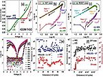
Partial-to-fully oxidized spectrum of Ti₃C₂Tₓ MXene-derived TiO₂ free-standing films for nonvolatile high endurance memristive data storage
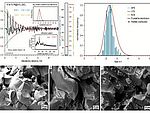
On-chip catalytic combustion of hydrogen using Pt and Ru quantum-crystallites on functionalized SiO₂ aerogels

Gas phase synthesis of mixed Cu₁.₈S-ZnS particles and the terminal phases in the reducing atmosphere
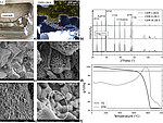
Reactive transport modelling of autogenous self-healing: Impact of portlandite content and degree of hydration

Controlled Synthesis of Copper Sulfide Nanoparticles in Oxygen-Deficient Conditions Using Flame Spray Pyrolysis (FSP) and Its Potential Application
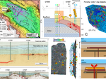
Sill Stacking in Subseafloor Unconsolidated Sediments and Control on Sustained Hydrothermal Systems: Evidence From IODP Drilling in the Guaymas Basin, Gulf of California
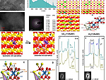
Stabilization of Ce³⁺ cations via U–Ce charge transfer in mixed oxides: consequences on the thermochemical water splitting to hydrogen
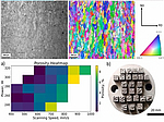
Making parts on Mars: Laser processing of iron contaminated by regolith simulant
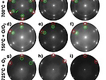
High-Temperature Growth of CeOₓ on Au(111) and Behavior under Reducing and Oxidizing Conditions
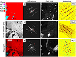
Effect of intrinsic heat treatment on the precipitate formation of X40CrMoV5–1 tool steel during laser-directed energy deposition: A coupled study of atom probe tomography and in situ synchrotron X-ray diffraction
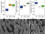
Towards coupling agent-free composites made from regenerated cellulose/HDPE by UV radiation-induced cross-linking
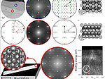
Hexagons on rectangles: Epitaxial graphene on Ru(1010)
2024
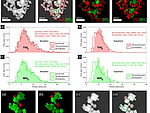
Characterization of structure and mixing in nanoparticle hetero-aggregates using convolutional neural networks: 3D-reconstruction versus 2D-projection
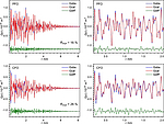
Synthesis, structural and spectroscopic characterization of defect-rich forsterite as a representative phase of Martian regolith

Photo Electrocatalytic Water Splitting Using Sn Doped In₂S₃ Homologous Series Synthesized in Oxygen Deficient Flame
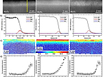
Composition and strain of the pseudomorphic α-phase intermediate layer at the Ga₂O₃/Al₂O₃ interface
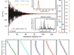
Mechanochemical synthesis of (Mg₁₋ₓFeₓ)₂SiO₄ olivine phases relevant to Martian regolith: structural and spectroscopic characterizations
2023
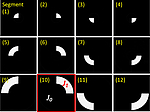
Sputter-Deposited β-Ga₂O₃ Films With Al Top Electrodes for Resistive Random Access Memory Technology
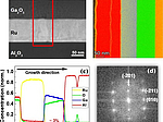
Growth and characterization of sputter-deposited Ga₂O₃ -based memristive devices
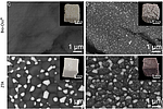
Gold Nanoparticle-Coated Bioceramics for Plasmonically Enhanced Molecule Detection via Surface-Enhanced Raman Scattering

Quantitative three-dimensional local order analysis of nanomaterials through electron diffraction

Timing of carbon uptake by oceanic crust determined by rock reactivity
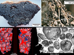
Dispersal of endolithic microorganisms in vesicular volcanic rock: Distribution, settlement and pathways revealed by 3D X-ray microscopy
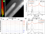
Correlative analysis on InGaN/GaN nanowires: structural and optical properties of self-assembled short-period superlattices

Low‐energy electron microscopy intensity–voltage data – Factorization, sparse sampling and classification

Oxygen Storage by Tin Oxide Monolayers on Pt₃Sn(111)

Halide-sodalites: thermal behavior at low temperatures and local deviations from the average structure
![[Translate to English:]](/fileadmin/_processed_/d/0/csm_XRM_Nils_e23e5d2a7b.png)
Gas Atomization of Duplex Stainless Steel Powder for Laser Powder Bed Fusion
2022
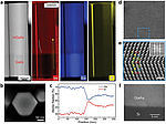
Deformed Honeycomb Lattices of InGaAs Nanowires Grown on Silicon-on-Insulator for Photonic Crystal Surface-Emitting Lasers
![[Translate to English:] [Translate to English:]](/fileadmin/_processed_/8/0/csm_FaltaFlege2022_4x3b_5f0c2b9289.png)
Growth Mechanism of Single-Domain Monolayer MoS₂ Nanosheets on Au(111) Revealed by In Situ Microscopy: Implications for Optoelectronics Applications
![[Translate to English:] [Translate to English:]](/fileadmin/_processed_/0/e/csm_Rosenauer2022_gold.jpeg_77341aa03b.png)
New Perspectives for Evaluating the Mass Transport in Porous Catalysts and Unfolding Macro- and Microkinetics
![[Translate to English:] [Translate to English:]](/fileadmin/_processed_/9/2/csm_Krause_2023_ac58139078.jpg)
Dose efficient annular bright field contrast with the ISTEM method: A proof of principle demonstration
![[Translate to English:] [Translate to English:]](/fileadmin/_processed_/b/7/csm_Gesing2022_red_147e86d8ed.jpeg)
On red tin (II) oxide: temperature-dependent structural, spectroscopic, and thermogravimetric properties
![[Translate to English:] [Translate to English:]](/fileadmin/_processed_/f/0/csm_Falta2022_4x3_02f7dddeec.png)
Phase Separation within Vanadium Oxide Islands under Reaction Conditions: Methanol Oxidation at Vanadium Oxide Films on Rh(111)

Effects of iron substitution and anti-site disorder on crystal structures, vibrational, optical and magnetic properties of double perovskites Sr₂(Fe₁₋ₓNiₓ)TeO₆
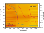
Nano-crystalline precursor formation, stability, and transformation to mullite-type visible-light photocatalysts
![[Translate to English:] [Translate to English:]](/fileadmin/_processed_/4/a/csm_Rosenauer2022_fee6db1bbe.jpeg)
Investigation of the dealloying front in partially corroded alloys

Enhanced weathering in the seabed: Rapid olivine dissolution and iron sulfide formation in submarine volcanic ash
![[Translate to English:] [Translate to English:]](/fileadmin/_processed_/e/6/csm_Iris_InGaN_image_Mapex_HP_01_e56fb752fa.png)
Angle-dependence of ADF-STEM intensities for chemical analysis of InGaN/GaN
![[Translate to English:] [Translate to English:]](/fileadmin/_processed_/7/3/csm_SiGe_experiment_120nm_21mrad_110ZA_VCA_EField_CoG_AllThicknesses_neu_4MAPEX_dee4066d55.png)
Towards the interpretation of a shift of the central beam in nano-beam electron diffraction as a change in mean inner potential
![[Translate to English:] [Translate to English:]](/fileadmin/_processed_/5/8/csm_Promoting_Effect_of_the_Residual_Silver_on_the_Electrocatalytic_Oxidation_of_Methanol_and_Its_Intermediates_on_Nanoporous_Gold_ecaae7ea01.png)
Promoting Effect of the Residual Silver on the Electrocatalytic Oxidation of Methanol and Its Intermediates on Nanoporous Gold
![[Translate to English:] [Translate to English:]](/fileadmin/_processed_/9/f/csm_Gesing2022png_92f1b590bc.png)
Revisiting the Growth of Large (Mg,Zr):SrGa₁₂O₁₉ Single Crystals: Core Formation and Its Impact on Structural Homogeneity Revealed by Correlative X-ray Imaging
![[Translate to English:] [Translate to English:]](/fileadmin/_processed_/a/0/csm_Open_Ceramics_2022_MurshedMaas_65ba8d3d7a.jpg)
Plasmonic porous ceramics based on zirconia-toughened alumina functionalized with silver nanoparticles for surface-enhanced Raman scattering
![[Translate to English:] [Translate to English:]](/fileadmin/_processed_/7/6/csm_2022_Wolpmann_et_al._2_51987c8d65.jpg)
Halide-sodalites: thermal expansion, decomposition and the Lindemann criterion
2021
![[Translate to English:] [Translate to English:]](/fileadmin/_processed_/3/c/csm_MurshedGesing2018_0b4edf1f65.png)
Structural, vibrational, thermal, and magnetic properties of mullite-type NdMnTiO₅ ceramic
![[Translate to English:] [Translate to English:]](/fileadmin/_processed_/6/8/csm_2021_Gogolin_et_al._1f9587172b.jpg)
Thermal anomalies and phase transitions in Pb₂Sc₂Si₂O₉ and Pb₂In₂Si₂O₉
![[Translate to English:] [Translate to English:]](/fileadmin/_processed_/1/2/csm_JPCC_FaltaJOK_2021_32bc60b000.gif)
Structural Transitions Driving Interface Pulses in Methanol Oxidation on Rh(110) and VOₓ/Rh(110): A LEEM Study
![[Translate to English:] picture](/fileadmin/_processed_/3/4/csm_2021_Luttge_et_al_5e8ece7690.jpg)
The role of crystal heterogeneity in alkali feldspar dissolution kinetics
![[Translate to English:] [Translate to English:]](/fileadmin/_processed_/8/4/csm_2021_Barreto_et_al._28744af394.jpg)
Influence of Processing Route on the Surface Reactivity of Cu₄₇Ti₃₃Zr₁₁Ni₆Sn₂Si₁ Metallic Glass

The Transition From MoS₂ Single-Layer to Bilayer Growth on the Au(111) Surface
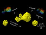
Quantitative 3D Characterization of Nanoporous Gold Nanoparticles by Transmission Electron Microscopy
![[Translate to English:] [Translate to English:]](/fileadmin/_processed_/7/9/csm_2021_Li_et_al._efc5cdd198.jpg)
Multiscale investigation of olivine (0 1 0) face dissolution from a surface control perspective

Precise measurement of the electron beam current in a TEM
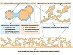
A review of contact force models between nanoparticles in agglomerates, aggregates, and films
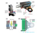
Angle-resolved STEM using an iris aperture: Scattering contributions and sources of error for the quantitative analysis in Si
![[Translate to English:] [Translate to English:]](/fileadmin/_processed_/7/8/csm_Dilissen_etal_2021_8d2c437aad.jpg)
Morphological transition during prograde olivine growth formed by high-pressure dehydration of antigorite-serpentinite to chlorite-harzburgite in a subduction setting
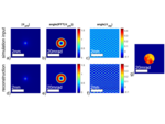
Accurate measurement of strain at interfaces in 4D-STEM: A comparison of various methods
2020
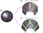
The morphology of VO₂/TiO₂(001): terraces, facets, and cracks
![[Translate to English:] [Translate to English:]](/fileadmin/_processed_/c/5/csm_Crystal_structure_of_KLi2Ho_BO3_2_4b94cad6c3.jpeg)
KLi₂RE(BO₃)₂ (RE = Dy, Ho, Er, Tm, Yb, and Y): Structural, Spectroscopic, And Thermogravimetric Studies on a Series of Mixed-Alkali Rare-Earth Orthoborates
![[Translate to English:] [Translate to English:]](/fileadmin/_processed_/5/a/csm_2020_Peschke_et_al._eee89995c9.jpg)
Crystal structure and temperature-dependent properties of Na₂H₄Ga₂GeO₈ – a novel gallogermanate
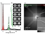
Influence of plasmon excitations on atomic-resolution quantitative 4D scanning transmission electron microscopy
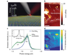
Imaging strain-localized excitons in nanoscale bubbles of monolayer WSe₂ at room temperature
![[Translate to English:] [Translate to English:]](/fileadmin/_processed_/9/d/csm_2020_Petersen_et_al._e2f2b38cb4.jpg)
On the nature of the phase transitions of aluminosilicate perrhenate sodalite
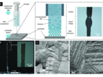
Strong Macroscale Supercrystalline Structures by 3D Printing Combined with Self‐Assembly of Ceramic Functionalized Nanoparticles
![[Translate to English:] [Translate to English:]](/fileadmin/_processed_/1/4/csm_2020_Schmidt_et_al._2a073e7bd1.jpg)
Adsorption of sulfur on Si(111)
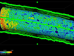
Crystal surface reactivity analysis using a combined approach of X-ray micro-computed tomography and vertical scanning interferometry
2019
![[Translate to English:] [Translate to English:]](/fileadmin/_processed_/7/d/csm_2019_Stapelfeldt_et_al._702bfd13a6.jpg)
Controlling the Multiscale Structure of Nanofibrous Fibrinogen Scaffolds for Wound Healing
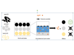
A new test system for unraveling the effects of soil components on the uptake and toxicity of silver nanoparticles (NM-300K) in simulated pore water
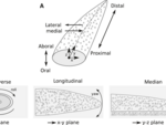
Morphology, shape variation and movement of skeletal elements in starfish (Asterias rubens)
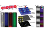
Electrical Polarization in AlN/GaN Nanodisks Measured by Momentum-Resolved 4D Scanning Transmission Electron Microscopy
![[Translate to English:] [Translate to English:]](/fileadmin/_processed_/a/9/csm_2019_Luttge_et_al._938d2bedd2.jpg)
Kinetic concepts for quantitative prediction of fluid-solid interactions

Influence of distortions of recorded diffraction patterns on strain analysis by nano-beam electron diffraction
2018
![[Translate to English:] [Translate to English:]](/fileadmin/_processed_/1/5/csm_2018_Heuer_et_al._fa59e11403.jpg)
Kinetics of pipeline steel corrosion studied by Raman spectroscopy-coupled vertical scanning interferometry
![[Translate to English:] [Translate to English:]](/fileadmin/_processed_/8/c/csm_2018_Robben_et_al._1980bc331f.jpg)
Low-temperature anharmonicity and symmetry breaking in the sodalite |Na₈I₂|[AlSiO₄]₆
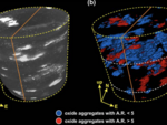
Textural evolution during high-pressure dehydration of serpentinite to peridotite and its relation to stress orientations and kinematics of subducting slabs: Insights from the Almirez ultramafic massif
![[Translate to English:] [Translate to English:]](/fileadmin/_processed_/6/3/csm_2018_Kirsch_et_al._bbc91c6013.jpg)
Temperature-dependent Structural and Spectroscopic Studies of (Bi₁₋ₓFeₓ)FeO₃
![[Translate to English:] [Translate to English:]](/fileadmin/_processed_/f/a/csm_2018_Lonescu_et_al._c2d1b01c52.jpg)
Discrimination of Ceramic Surface Finishing by Vertical Scanning Interferometry

Ambient occlusion – A powerful algorithm to segment shell and skeletal intrapores in computed tomography data
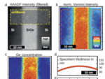
Quantitative HAADF STEM of SiGe in presence of amorphous surface layers from FIB preparation
2017
![[Translate to English:] [Translate to English:]](/fileadmin/_processed_/0/c/csm_2017_Teck_et_al._2ba5815a43.jpg)
Structural and spectroscopic comparison between polycrystalline, nanocrystalline and quantum dot visible light photo-catalyst Bi₂WO₆
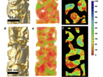
Quantitative determination of residual silver distribution in nanoporous gold and its influence on structure and catalytic performance
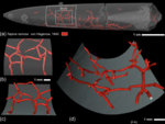
Classical and new bioerosion trace fossils in Cretaceous belemnite guards characterised via micro-CT
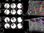
3-D microstructure of olivine in complex geological materials reconstructed by correlative X-ray µ-CT and EBSD analyses
2016
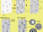
Reaction-induced porosity and onset of low-temperature carbonation in abyssal peridotites: insights from 3D high-resolution microtomography
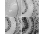
Growth and structure of ultrathin praseodymium oxide layers on ruthenium (0001)
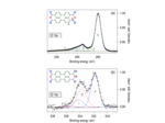
Isotropic thin PTCDA films on GaN(0001)


